Malek est un petit garçon âgé de 4 ans. Il est bon état, très actif, mais lors de la miction une petite tuméfaction kystique sur la face ventrale de la verge apparaît. Elle augmente de volume et prend l’aspect d’une « ampoule » , elle n’est ni douloureuse ni inflammatoire.
Le problème avec cette malformation c’est que la miction n’est jamais totale. La vidange progressive de la tuméfaction tache les sous-vêtements de Malek et donne l’impression d’une incontinence urinaire : “ne pas pouvoir retenir ses urines”.
Croyant, que Malek fait exprès de souiller son slip d’urines, il est pratiquement puni après chaque miction.


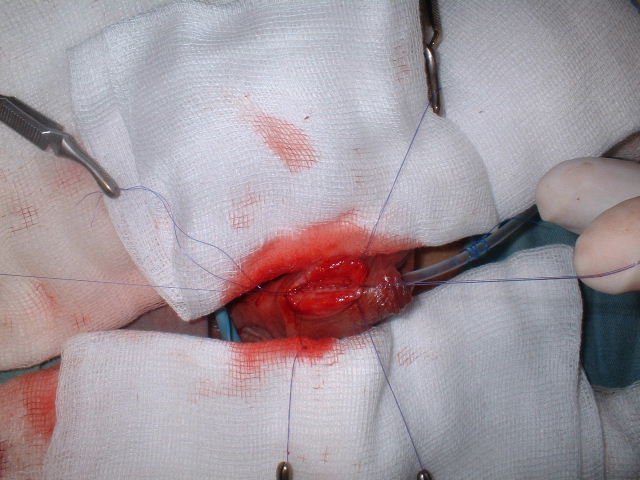
Malek est opéré, il a eu une résection de cette «urètrocèle» et une urètroplastie. Depuis il va bien.
Pour plus de renseignements:
1/ http://mesannexes01.unblog.fr/files/2011/03/diverticuledeluretreantrieur.pdf
2/ Primary anterior urethral diverticulum via M Sailukar, K Parikh, V Phadke, N Chakrabarti
Diverticula of the male urethra can be classified as congenital or acquired. Congenital diverticula of the anterior urethra are very rare and typically occur at the penoscrotal junction. They are usually wide-mouthed and the distal edge may act as a valve that can obstruct the urine flow.[3] There are very few cases of giant diverticulum which are reported.[4],[5] In our case, a giant primary diverticulum was present in the distal penile urethra.
Etiological factors in congenital diverticula have been summarized by Williams and Retik and include intrauterine distal urethral stenosis; lesser degree hypospadiasis or congenital cystic dilatation of the normal or accessory periurethral glands.[6]
These anomalies are rare with no genetic pre-disposition. Anterior urethral valves associated with diverticulum are 10 times less common than posterior urethral valves while anterior urethral valves alone or diverticulum alone is 25-30% less frequent. The clinical presentation depends upon the age and degree of presentation. In neonatal age and infancy, symptoms related to urinary infection predominate while in older children voiding problems are the presenting complaints.
The anatomical interpretation of these lesions is variable; some authors combine all these abnormalities under the term 'Diverticula'. Others only refer 'Anterior urethral valves' considering that diverticula and anterior urethral valves represent the same pathology. Nevertheless, many authors clearly distinguish valves and diverticula.[7] Usually the diverticula are saccular, communicating with urethral lumen. They are present on the ventral aspect of the urethra and are variable in size. With the stagnation of urine, stones can be formed inside the diverticulum making it a unique presentation.[8]
The diagnosis depends essentially on voiding cystourethrography, which must opacify the whole of the urethra. Retrograde urethrography is sometimes needed in case of a giant diverticulum. Though associated anomalies are rare, it is always advisable to look for them by doing additional investigations. Vesicourethral reflux,[9] hydronephrosis, Prune-belly syndrome and posterior urethral valves have been reported in literature. Whereas, many authors have used the term anterior urethral valves and anterior urethral diverticula synonymously, we feel that there is a need to differentiate the two because of the fact that the treatment differs for both conditions. Whereas, primary valves can be successfully treated by transurethral endoscopic resection,[10] in case of primary diverticula, open resection with or without cystostomy is necessary.
Aucun commentaire:
Enregistrer un commentaire